|
|
|
|
International Symposium on Plastination 07/21/07 |
| brochure | |||
|
|
Lecture 2
Principles of Plastination
Prof.Dr. Aly Eldeen Abd Elbasset Aly Project Manager of ETAP, Plastination Laboratory, Faculty of Veterinary Medicine, Zagazig University ,Egypt
Introduction The biological materials after death is decomposed by autolysis and putrefaction. Since the beginning of our life, trials have been made to keep the body in normal shape, to keep the mortal frame for coming back to life later e.g. Egyptian mummification 6000 B.C. and cryo-preservation in modern life. Egyptians successfully preserved their bodies via a process called mummification. Mummification dried out the body, using a substance called natron. Fixation by formalin was discovered by Blum 1896, which protect the cadavers against deterioration. Disadvantage of formalin include smell, shrinkage and discoloration of the tissue. Formalin, however, is still mainly used for preservation in Anatomy Department because of its price and preservation properties. In 1978 Gunther von Hagens invented Plastination in Heidelberg University , Germany. What is Plastination? - A method of preserving anatomical specimens by replacing the tissue water and lipid with a curable plastic polymer. The specimens preserved in this manner are permanent, clean, non-toxic and dry.
Why is it useful in Anatomy? - Plastinated specimens retain textures and structures of tissue and are therefore an invaluable teaching resource in anatomy. Plastinated specimens have none of the usual hazards and restrictions associated with the study of anatomical specimens e.g. use of gloves, toxic fumes, contagions etc, and are more robust than the original specimen.
Theory and Procedures (Von Hagens G. 1985) In Plastination process, water and lipids in biological tissues are replaced by curable polymers (silicone, epoxy, polyester) which are subsequently hardened, resulting in dry, odorless and durable specimens. The class of polymer used determines the optical (transparent or opaque) and mechanical (flexible or firm) properties of the impregnated specimen.
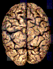
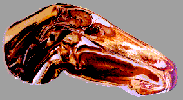
Silicone 10 is used for whole specimens and thick body and organ slices to obtain a natural look.
Epoxy resins are used for thin, transparent body and organ slices.
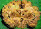
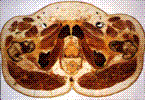
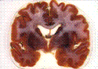
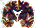
Polyester-copolymer (P 40) is exclusively used for brain slices to gain an excellent distinction of gray and white matter.
The technique consists of four main steps:
Fixation can be done by almost all conventional fixatives (e.g. formalin). Dehydration is achieved mainly by acetone because acetone also serves as the intermediary solvent during impregnation. Forced impregnation is the central step in plastination: vacuum forces the acetone out of and the polymer into the specimen. Hardening (Curing): Finally the impregnated specimen is hardened by exposing it to a gaseous hardener (silicone), or by UVA-light and heat (polyester, epoxy). Plastinated specimens are perfect for teaching, particularly for neuroanatomy. Silicone plastinated brains are useful because they can be grasped literally and they are almost everlasting. Polyester plastination of brain slices provides an excellent distinction of gray and white matter and thus a better orientation. Plastination is carried out in many institutions worldwide and has obtained great acceptance particularly because of the durability, the possibility for direct comparison to CT- and MR-images, and the high teaching value plastinated specimens have. If a university takes the decision and commitment to help this project the results will be excellent and useful for new students who will shape the future in research,clinical knowledge and teaching. References: 1-Aly Eldeen Abd Elbasset Aly,Alexander Probst, Mircea-Constantin Sora und Horst Erich König ( Oktober ,2005 ).Plastination-reale Abbilder Lebender Structuren. Zeitschrift der Veterinärmedizinischen Univ. Wien,Heft 03/05 2-Aly Eldeen Abd Elbasset Aly Establishment of Zagazig Plastination Laboratory at Faculty of Veterinary Medicine,Zagazig University: Abstract book of the 29th Scientific Conference of The Egyptian Anatomical Society ,Ein Shams University (December 2005). 3- A.E.Basset Aly, , Mervat M.H. Konsowa and S.Abdel Aziz : Enhancing Teaching of Anatomy of The Head of Goat by Plastinated Specimens. Abstract book of the 29th Scientific Conference of The Egyptian Anatomical Society, Ein Shams University (December 2005). 4-Blum 1896: Cited by Weiglein, AH.2001: Preservation of Biological Tissue: Yesterday-Today- Tomorrow.J.Int.Soc.Plastination16:31-41, 2001. 6-Von Hagens G. 1985: Heidelberg Plastination Folder: Collection of all technical Leaflets for Plastination.Heidelberg,Germany:Anatomische Institut . 1,Universitat Heidelberg. 7-Weiglein ,A. 2004 : Seminar Plastination. Skopje 3-8 May 2004.
|
||
This site was last updated 08/10/06